
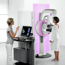
What is Mammography?
It is a specialized X Ray of the breasts with low voltage and high ampere beams. Breasts are compressed between the plates. There are two standard views – Crainocaudal and Mediolateral. Additional views are taken if required.
It is an X ray with low dose of radiation, hence no risk of Malignancy.
It should be done so that lesions are detected at a very early stage when they are not even clinically palpable and at a stage when the disease is curable.
It can differentiate between benign(non-cancerous) and malignant(cancerous) breast lumps.
Mammography
- 40-50 years – every 1-2 years, as per doctors advice
- 35-40years – baseline mammogram
- 50 years–every year
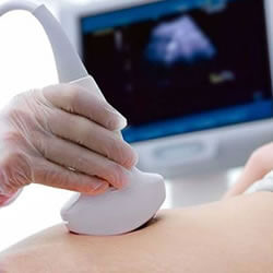
Ultrasonography
Ultrasonography is usually combined with mammography as a protocol now. It detects small lumps and cysts at an early stage. It is also useful to take biopsy- FNAC and Core Biopsy. It is also combined with elastography.
A newer technique called Vaccum Assisted Biopsy (VAB) is a very safe and minimally invasive procedure, wherein the breast tissue is taken and sent for biopsy. Even a very small lump, which is not palpable clinically, but seen under Breast imaging, can be removed and biopsied.
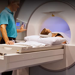
MRI (MR Mammogram)
MR Mammogram is done for Characterization of an indeterminate lesion after a full assessment with mammography, ultrasonography, and physical examination. It is advised for evaluation of suspected multifocal or bilateral lesions. It is done in women with high risk and genetic predisposition.
- PET SCAN
